
Articles
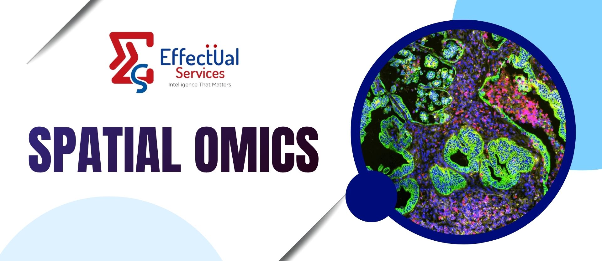
Spatial Omics
Spatial Omics
In the field of omics, spatial biology is expected to be the "next big thing." It offers to raise an ambitious idea the localization of tissue components at the cellular and molecular levels—on the strength of new technology. Though the idea has existed for many years, advances in spatial biological technology are bringing the concept to new heights. These technologies do, in fact, hold the potential to bring in a new age in omics.
The phrase "spatial omics" refers to a group of technologies that enable -omics data to be superimposed on tissue pictures. These methods thus offer information on the spatial localization of the identified subpopulations within the tissue of origin, as well as their proximity to the extracellular matrix, blood vessels, and other tissue components, in addition to the identification of molecular subpopulations of cells.
The history of contemporary spatial approaches was retraced by Lior Pachter, PhD, a computational biology professor at the California Institute of Technology, and his doctoral student Lambda Moses in a recent Nature Methods review titled "Museum of Spatial Transcriptomics" (2022; 19: 534–546). Around that time, a technique for radioactively in situ hybridizing ribosomal RNA was introduced. Numerous spatial techniques have since been developed.
Currently, a number of methods are being developed for the study of RNA transcripts or proteins in the spatial context of tissues. Furthermore, over a dozen companies—including 10x Genomics, Akoya Biosciences, Lunaphore Technologies, Bruker (formerly NanoString Technologies), Rebus Biosystems, Resolve Biosciences, Veranome Biosystems, and Vizgen—are pursuing the commercialization of these methodologies. These businesses are producing tools and making their own claims and forecasts about the capabilities of their platforms.
It's evident that more businesses are promoting spatial technology and that more scholars are including spatial experiments in their research reports and funding requests. Consequently, it's critical to hear from the scientists working at the edge of space. What these researchers have to say about the current state and potential future directions of spatial biology.
Why does spatial have such power?
Compared to single cell analysis, spatial allows one to better grasp how cell-type compositions are changing within a context. It is anticipated that spatial methods will be particularly useful for describing cellular connections and enhancing our knowledge of gene-level intercellular communications.
With the recent release of Molecular Cartography, a technology from Resolve Biosciences, scientists can now view subcellular gene expression activities at high resolution. Additionally, Resolve stated that a number of prestigious research institutes had already deployed the platform. A couple of these establishments are VIB-KU Leuven. There, in the perivascular niche microenvironment of a mouse primary melanoma tumor, the technology assisted researchers under the direction of Christophe Marine, PhD, in partially resolving different cell populations.
In the field of omics, spatial biology is expected to be the "next big thing." It offers to raise an ambitious idea—the localization of tissue components at the cellular and molecular levels—on the strength of new technology. Though the idea has existed for many years, advances in spatial biological technology are bringing the concept to new heights. These technologies do, in fact, hold the potential to bring in a new age in omics.
Currently, a number of methods are being developed for the study of RNA transcripts or proteins in the spatial context of tissues. Furthermore, over a dozen companies—including 10x Genomics, Akoya Biosciences, Lunaphore Technologies, Bruker (formerly NanoString Technologies), Rebus Biosystems, Resolve Biosciences, Veranome Biosystems, and Vizgen—are pursuing the commercialization of these methodologies. These businesses are producing tools and making their own claims and forecasts about the capabilities of their platforms.
Spatial Omics Technologies
Laser capture microdissection-based approaches
The earliest efforts to disseminate the high-throughput spatial tissue structure were attributed to techniques based on laser capture microdissection (LCM). With this method, tissues are divided into tiny segments using LCM, and these segments are then profiled using high-throughput technologies like RNA-seq. For instance, LCM-seq follows the spatial transcriptome at the single-cell level by integrating LCM with single-cell RNA-seq (scRNA-seq). This method enables the precise measurement of tissue structural compartments and the identification of diverse cell subpopulation distributions within tissues. Topographic single-cell sequencing (TSCS) aims to spatially record the genomic copy number profile of individual tumor cells by a similar approach. The results demonstrate a direct genomic lineage of breast cancer cell development by using TSCS in breast cancer samples. It's interesting to note that the majority of mutations and copy number aberrations were present before invasion, according to the authors, suggesting that cancer cells are ready to spread before they do. This method enables the objective identification of 2D copy number changes. Sequencing of geographic positions (GEO-seq). Using a comparatively small number of cells, a different technique combining the two technologies (scRNA-seq and LCM) can capture the spatial transcriptome. Nonetheless, each site in GEO-seq captures more cells than in LCM-seq. In a similar vein, Tomo-seq permits RNA-seq on specific sections and cryosectioning of the region of interest.
Image-based in situ transcriptomics
With image-based in situ transcriptomics technology, the spatial architecture of tissue transcripts may also be captured. Multi-RNA detection is possible with single-molecule fluorescence in situ hybridization (smFISH). Ouroboros single-molecule FISH (osmFISH), expansion FISH (ExFISH), and sequential FISH+ (seqFISH+) are based on smFISH and are intended to enhance the number of discovered genes (up to 10,000). In 2019, the gene throughput was elevated to approximately 100,000 thanks to the reliable single-molecule imaging method known as multiplexed error-robust fluorescence in situ hybridization (MERFISH), which can detect between 100 and 1000 different RNA species in hundreds of individual cells. Similarly, 160–1020 genes are tested to be concurrently captured by Spatially-resolved Transcript Amplicon Readout mapping (STARmap), which combines hydrogel-tissue chemistry, tailored signal amplification, and in situ sequencing.
Certain RNAs that are abundant in cellular compartments or even high-order chromatin structure have been found by image-based approaches. To put it briefly, the majority of image-based in situ transcriptomics methods are unable to capture the transcriptome profile in its entirety, but they do provide single-cell or even subcellular resolution within tissues, which makes it possible to identify the intricate biological states of cancer cells.
Spatial barcoding-based transcriptomics
Spatial barcode-based methods, as opposed to image-based in situ transcriptomics, enable the unbiased sequencing of RNA species at the transcriptome level. One of the most popular spatial omics technologies, spatial transcriptomics (ST, also known as Visium), allows the sequencing of 6 mm × 6 mm tissues with each spot at the resolution of approximately 100 μm containing 2–10 cells. 10x Genomics has since launched Visium HD (2024), enabling whole-transcriptome spatial discovery at near single cell-scale resolution, representing a major advancement over the original Visium platform.
The ability to extract thousands of genes from formalin-fixed paraffin embedding (FFPE) tissues and those with low-level transcript expression is one of the benefits of this technique.
Then, with a resolution of 2 μm, an improved technique called high-definition spatial transcriptomics (HDST) was created. This method paves the way for single-cell resolution spatial measurement of intricate tissues. Another high-resolution spatial sequencing technologies, Slide-seq and Slide-seqV2, can successfully characterize the spatiotemporal developmental trajectory of the mouse neocortex and achieve ~50% RNA capture efficiency of scRNA-seq. Improved Spatio-Temporal Resolution By utilizing a combination of in situ RNA capture and DNA nanoball chips, omics-sequencing (Stereo-seq) may achieve a resolution of around 0.5 μm for each bin. The recently created Seq-Scope method for spatial transcriptome sequencing can also achieve sub-cellular resolution (0.5–1 μm). It is based on solid-phase amplification of randomly barcoded single-molecule oligonucleotides. These techniques provide us a distinct advantage in investigating novel roles for organelles and could result in a significant breakthrough in our comprehension of spatiotemporal molecular medicine.
Spatial proteomics
Numerous proteins can now be detected without losing their spatial location thanks to the rapid advancements in spatial proteomics. A highly multiplex method to capture the spatial intensity of proteins is mass spectrometry-based. Multiplexed ion beam imaging (MIBI) may analyze one hundred markers of the same tissue by imaging labeled antibodies using secondary ion mass spectrometry.
With the use of this technology, immune cell subpopulations and their spatial patterns inside tumors may be precisely quantified. IMC allows the imaging of more than 100 antibodies and is also reliant on metal-tagged antibodies. This approach provides previously unheard-of possibilities to investigate the topological function units of TME and the composition of regional immunity.
A different technique called CO detection by indexing (CODEX) uses imaging antibodies linked to barcodes to profile up to 50 proteins on a single slide. All those techniques are, however, somewhat expensive and rely on the effectiveness of antibodies. Increasing the current throughput to proteome-wide remains difficult. Instead of identifying useful proteins from the proteomics data, bias can possibly have been present during the panel of markers' construction. To fully comprehend cancer systems and interpret intratumor heterogeneity, it is imperative to discern the variations in metabolites and the spatial arrangement of tissues.
Nevertheless, little is known about the spatial metabolic characteristics of tumors. Metabolite detection is made possible with matrix-assisted laser desorption ionization imaging mass spectrometry (MALDI-IMS) without sacrificing spatial information.
An additional technique to identify the spatial dynamics metabolites without damaging the tissues is desorption electrospray ionization (DESI)-IMS.
The multiplex capability is further increased using airflow-assisted desorption electrospray ionization (AFADESI)-IMS, which can cover 1,500 metabolites.
Using this technique to profile esophageal squamous cell carcinoma revealed the spatial small structure of some metabolites, such as uridine phosphorylase 1 (UPase1) and pyrroline-5-carboxylate reductase 2 (PYCR2).
Spatial metabolomics will be a valuable tool in the emerging field of spatial omics for the identification of new disease signatures.
Novel approaches to probe cell heterogeneity and extracellular matrix biology
Cellular processes are significantly impacted by biomolecular and biomechanical inputs from the extracellular matrix (ECM), as well as complex intercellular connections.
By identifying distinct cellular subpopulations or microenvironmental signatures typical of normal or pathological tissues, traditional transcriptomic and proteomic approaches have shed light on the progression of disease. However, these methods do not investigate the relationship between a given cellular state and its interactions with neighboring cells or its surrounding extracellular matrix (ECM) with multiparametric characterization (i.e. ECM alignment, mechanical forces, crosslinking, etc.).
New developments in spatial-omic techniques can offer direct tissue mapping of expression patterns with great precision, akin to that of mass spectroscopy and scRNA-seq.
Spatial-omics has the potential to transform ECM research and our understanding of fibrotic diseases by allowing us to examine hitherto undiscovered signaling modalities because it can maintain the spatial context of cells within samples, their cellular geometry, and their surrounding ECM.
The Market
The market for spatial omics solutions was estimated to be worth $251 million in 2022. Updated market analyses now value the global spatial omics market at approximately $397–587 million in 2024, reflecting faster-than-projected growth. The market is projected to grow at a CAGR of approximately 10–13% to reach $830 million to $1.23 billion by 2030–2034, driven by technological advancements, rising oncology applications, and increased adoption in precision medicine.
Market segmentation
Instruments, kits, consumables, and services are the different product types.
Diagnostics, translation studies, drug development and discovery, single-cell analysis, and cell biology are a few examples of application areas.
Proteins, RNA, and DNA are among the samples that are examined.
End consumers include academic and research institutions, biopharma and biotech firms, and Contract Research Organizations (CROs).
The SignalStar™ Multiplex IHC technology was introduced by Cell Signaling Technology. This is a new method for spatial biology research that labels up to eight targets in FFPE tissues concurrently with flexible, fully validated antibody panels using high-throughput, mid-plex IHC experiments. The optimized, ready-to-use panels can be utilized to investigate biomarker coexpression patterns, cellular presence, location, and function right out of the box.
The ground-breaking DBiT-seq platform, among other innovative spatial epigenomic technologies, is provided by AtlasXomics. The DBiT-seq discovery platform is a hybrid of next-generation sequencing and microfluidics. Multi-omic probes (for mRNA, genomic DNA, or proteins, for example) are first added directly to a tissue piece to begin the procedure. Microfluidic chips are applied sequentially and orthogonally to deliver x- and y-barcodes, forming an in situ x-y coordinate system in tissue. Every point where a microfluidic row and column cross produces a TIXEL™, a square-shaped piece of tissue. After the spatially tagged analytes in each TIXEL™ are extracted and sequenced, a multi-omic map at cellular resolution is created using the generated data.
With the introduction of its revolutionary DNA chip technology, Centrillion Tech. Their novel wafer-scale produced high-resolution spatial DNA chips, which leverage light-directed synthesis and semiconductor manufacturing, allow for high-resolution spatial transcriptomics study. These chips can be customized to study lengthy polymers like chromosomal DNA and to take on a wide range of spatial biology concerns.
In 2020, Rarecyte entered the field of spatial biology with the release of their Orion™ Multiplex Imaging Instrument. Whole Slide Multiplex Immunofluorescence Imaging provides biomarker depth and versatility in a single stain and scan phase, unlocking high-plex spatial biology. In just a few hours, it can provide thorough phenotypic profiling and characterization of tissue architecture, tumor heterogeneity, and the immune response for entire sections thanks to its speed and resolution combination.
Up to 60 proteins can be seen and described in a single formalin-fixed, paraffin-embedded (FFPE) 4 μm tissue segment thanks to Neogenomics' MultiOmyx™ technology. Utilizing the MultiOmyx™ multiplexed IF staining procedure, slides are prepared and stained. Conjugated fluorescent antibodies are added to the slide for each staining cycle, and then the stained slides are imaged. After the dye is removed, another round of staining with a different pair of fluorescent antibodies is possible.
A Swedish biotech business called Navinci Diagnostics specializes in cutting-edge proximity ligation assay-based spatial proteomics solutions. Protein localization, post-translational changes, and protein-protein interactions can all be seen and measured with the Naveni™ Proximity Ligation Technology. To improve specificity and sensitivity, the method relies on dual antibody recognition and signal amplification. These days, the technology is used in five product categories: target-specific products like Naveni PTMs (post-translational modifications) and Naveni PPI (protein-protein interaction), and flexible products like NaveniFlex, Naveni TriFlex, and NaveniBright.
Large biotech company Standard BioTools was formerly known as Fluidigm. They provide single-cell insights with their Hyperion™ Imaging System, which is driven by Imaging Mass Cytometry™. A greater comprehension of the complex spatial interactions between cells, pathways, and phenotypes inside the tissue microenvironment is one aspect of this. At subcellular resolution, the device is capable of simultaneously examining over 40 markers.
STOmics was founded in 2020, and their patented Stereo-seq (SpaTial Enhanced REsolution Omics-sequencing) technology allows for the in situ sequencing of tissues, enabling a solid research foundation for a thorough understanding of the relationship between gene expression, cell morphology, and cellular microenvironments. The technology captures mRNA within the tissue on the chip and restores cellular spatial location through spatial coordination barcoding. It allows for simultaneous transcriptome analysis and research from fresh frozen tissue at the molecular, cellular, subcellular, and tissue levels.
With its patented DNBSEQ technology, Complete Genomics offers actual PCR-free NGS that is incredibly precise and efficient. The DNBSEQ-T7 Sequencer is designed to maximize productivity and efficiency in big sequencing projects, including single-cell omics, spatial omics, deep exome, transcriptome, and whole genome sequencing. Spatial omics applications are also possible with their G400 device.
An early-stage startup, Steam Instruments is creating and commercializing technology that enhance mass spectrometry's spatial resolution, quantum efficiency, and mass range. With the ability to detect nearly any mass ion or biomolecule, they are creating quantum ion detectors and quantum ion imaging detectors. They anticipate making unprecedented rapid advances in the fields of high-resolution nuclear forensics mapping, ultra-high-mass biomolecule analysis, and direct-from-tissue biomolecule mapping and quantification. These advances will be complemented by their innovative ion imaging optics and high-mass spectrometer designs.
Within this industry, 10x Genomics stands as one of the biggest companies. They provide in-situ, spatial, and single-cell technology solutions. Their next-generation molecular profiling tool, Visium Spatial Gene Expression, allows tissue classification based on total mRNA. In 2024, 10x Genomics launched Visium HD, which enables whole-transcriptome spatial discovery at near single cell-scale resolution — a significant advancement over the original Visium platform. The company also launched the Xenium Prime 5K panel in June 2024, enabling measurement of 5,000 genes with industry-leading speed and throughput.
Bruker (formerly NanoString Technologies): NanoString Technologies, which had created products based on a revolutionary digital molecular barcoding technique originally developed at ISB under the guidance of Dr. Leroy Hood, filed for Chapter 11 bankruptcy in February 2024 following a patent infringement case. Its assets were subsequently acquired by Bruker Corporation in May 2024 for approximately $392.6 million. Bruker now operates the nCounter® Analysis System, the GeoMx® Digital Spatial Profiler (DSP), and the CosMx™ Spatial Molecular Imager (SMI) platform under the NanoString brand, continuing to allow scientists to visualize molecular interactions in three dimensions.
Vizgen (now merged with Ultivue as of October 2024, operating under the Vizgen name) created a spatial transcriptomics technology called MERFISH (Multiplexed Error-Robust Fluorescence in situ Hybridization), which allows for the high accuracy and unparalleled detection efficiency of imaging RNA molecules at subcellular resolution.
In order to meet the varied needs of researchers in discovery, translational, and clinical research, Akoya Biosciences provides a comprehensive range of spatial phenotyping solutions. These solutions enable researchers to phenotype cells with spatial context and visualize how they organize and interact to influence disease progression and response to therapy. Products include PhenoImager HT Instruments, PhenoCycler®, PhenoCode™ Panels, and PhenoImager® Fusion.
Ultivue merged with Vizgen in October 2024, creating the largest private spatial biology company, operating under the Vizgen name. The combined entity unites Vizgen's breakthrough MERFISH spatial genomics technology with Ultivue's high-fidelity InSituPlex® assays and AI-powered STARVUE™ platform, delivering a unified solution for spatial multi-omics research.
A Bruker firm, Canopy Biosciences uses its own technology, ChipCytometry™, a quantitative multiplex immunofluorescence imaging technique, to map cell populations at single-cell resolution and analyze complex tissues spatially.
With the use of their AI-enabled cTA® pathology platform, Flagship Biosciences, a technology-driven provider of biomarker services and spatial biology, is able to provide the most precise and educational data out there.
The MIBI™ (multiplexed ion beam imaging) platform, created by Ionpath, overcomes the drawbacks of conventional IHC and provides highly multiplexed, quantitative single-cell analysis to enable a deeper understanding of the tissue microenvironment.
A significant multinational producer of life sciences is Miltenyi Biotec. With its sophisticated protein multiplexing capabilities, the MACSima™ Platform, driven by MICS (MACSima Imaging Cyclic Staining) technology, allows researchers to completely investigate spatial biology. The UltraMicroscope Blaze™ Platform enables cellular-level automated 3D imaging.
Lunaphore, acquired by Bio-Techne in July 2023, develops fully automated spatial biology solutions that turn any assay into multiplex spatial biology with an easy-to-use methodology. Based on a revolutionary chip technology that can extract spatial proteomic and transcriptomic data from tumors and other tissues, Lunaphore's solutions are now integrated into Bio-Techne's broader spatial biology portfolio.
The Rebus Esper, the first instrument made by Rebus Biosystems, is a fully automated, fully integrated spatial omics platform that provides quantitative, subcellularly resolved single-molecule, single-cell data. Combining on-system chemistry, intuitive software, and advanced imaging has resulted in an end-to-end solution that requires little manual labor.
The new GenePS device from Spatial Genomics, Inc. makes it simple to photograph and decode complicated molecular identities and locations directly within single cells and intact tissue microenvironments. It does this by using Sequential Fluorescence in Situ Hybridization, or seqFISH.
The only spatial omics tool that uses semiconductor industry imaging and process control technologies to create a stable and dependable platform for implementing complex spatial biology workflows is Veranome Biosystems' Spatial Analyzer (Model VSA-1).
The initial product from Curio Bioscience is the only continuous high-resolution spatial whole-transcriptome solution that can be used with the current DNA sequencing infrastructure to map gene expression from fresh frozen tissues in an unbiased manner.
The RNAscope™ ISH, a multiplex fluorescent and chromogenic in situ hybridization method, is a groundbreaking tool available from Advanced Cell Diagnostics. By amplifying target-specific signals in situ, ACD's proprietary probe design facilitates the identification and measurement of individual RNA molecules, yielding spatial gene expression data.
A technology developed by Acuity Spatial Genomics, a Bruker subsidiary, allows for the simultaneous display of the entire 3D genome in a single cell and offers up new avenues for spatial multiomic analysis for discovery. The company's OligoFISSEQ and OligoFISSEQ HD technologies, which are based on proprietary technologies created at Harvard University, enable researchers to see the 3D chromatin architecture and genome organization of individual cells right in situ.
Alpine Glow Scientific offers a workflow from sample preparation to visualization with 3D analysis as part of their end-to-end solution. Using an open top light-sheet (OTLS) microscope with patented geometry, they can analyze whole tissue samples or traditional multi-well plates with simplicity thanks to its user-friendly design. High throughput analysis is facilitated by this fully automated system, and complicated spatial biology applications can be seen and quantified in three dimensions by researchers using cloud computing and AI-enabled software.
Please feel free to reach out to one of our subject matter experts at info@effectualservices.com to explore how we can help you & make the world a better place to live in!!!
About Effectual Services
Effectual's SPATIAL OMICS RESEARCH FRAMEWORK is a deep dive into this ecosystem and shall help you understand the intricacies of this nascent innovative domain with insights backed with credible data sources. Some ways we can help include, but not limited to - Performing any previous art or freedom to operate searches to help you better grasp the environment surrounding your invention or business endeavors. If certain methods of IP protection are more appropriate for your technological or business goals, we can help you strategize effectively to plan for future & in making continuous innovation a part of your working model.
Solutions Driving Innovation & Intelligence


Enabling Fortune 500's, R&D Giants, Law firms, Universities, Research institutes & SME's Around The Globe Gather Intelligence That
Protects and Nurtures Innovation Through a Team of 250+ Techno Legal Professionals.